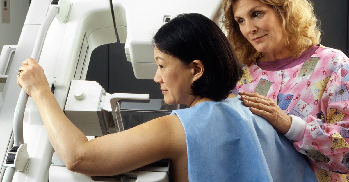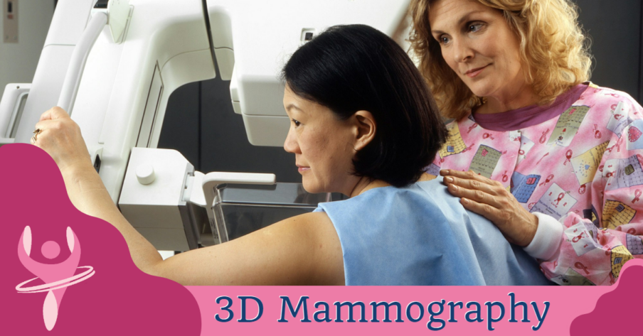
Capitol Imaging Services offers the advanced technology of 3D mammography within a relaxed non-hospital environment. 3D mammography, also known as breast tomosynthesis, was approved by the Food and Drug Administration (FDA) in 2011. Radiologists now have the ability to view inside the breast layer by layer, helping to see the fine details more clearly by minimizing overlapping tissue.
During a 3D mammogram, multiple low-dose images known as “slices” of the breast are acquired at different angles. With 3D technology, the radiologist can view a mammogram in a way never before possible.
The 3D exam is a separate procedure that is performed at the same time as your regular mammogram. Radiologists also use computer aided detection (CAD) software, along with 3D mammography, for a greater ability to find cancer at its earliest stages.
When would I get a 3D Mammography?
Breast cancer screening mammograms are often recommended on an annual basis, usually performed one year and one day after the previous screening. Annual screenings mammograms have been shown to help reduce the number of deaths from breast cancer among women ages 40 to 70.
Mammograms are also recommended for younger women who have symptoms of breast cancer or who have a high risk of the disease.
Women and their medical providers often prefer 3D mammography because it has been proven to provide results in superior testing performance in critical areas such as:
- EARLIER DETECTION by minimizing the impact of overlapping breast tissue, 3D mammography can help improve breast cancer screening and detection. With 3D mammography, studies indicate radiologists are currently experiencing up to a 30 percent increase in breast cancer detection.
- FEWER CALLBACKS as 3D mammography helps distinguish harmless abnormalities from real cancers, leading to fewer callbacks and less anxiety for women. With 3D mammography, radiologists may be able to reduce patient callback rates by as much as 20 to 30 percent.
- BETTER VISUALIZATION as radiologists can better see the size, shape and location of an abnormality.
What Will I Experience?
Our technologist will position your breast in the mammography unit. Your breast will be placed on a special platform and compressed with a clear plastic paddle. The technologist will gradually compress your breast.
You will be asked to change positions between images. The routine views are a top-to-bottom view and an angled side view. The process will be repeated for the other breast.
You will feel pressure on your breast as it is squeezed by the compression paddle. Some women with sensitive breasts may experience discomfort.
There are a variety of reasons that breast compression is necessary for a mammogram. Breast thickness needs to be evened out so that all tissue can be visualized. Spreading out breast tissue will minimize the likelihood that small abnormalities will be hidden by overlying breast tissue. Compression holds the breast in place to minimize any blurring that could result from motion.
A screening 3D mammogram will take approximately 10 to 12 minutes to complete. A diagnostic mammogram may take up to 30-45 minutes to complete.