

A ganglioglioma is a rare type of brain tumor. Tumors are issues that grow more rapidly than normal. Gangliogliomas are types of brain tumors that are made of mixed groups of cells. A cell is the smallest, most basic unit of life, that is capable of existing by itself. Gangliogliomas are partly made of abnormal glial cells. Glial cells are cells that support and maintain other cells. Gangliogliomas are also partly made of neurons (nerve cells) that are in various degrees of abnormality. Whereas neurons transmit and receive information to and from one another, glial cells do not.
Supportive tissue known as stroma, which contains fibers and blood vessels, are also found in gangliogliomas. Fibers are flexible, threadlike objects found outside of cells. Blood vessels are tube shaped structures that carry blood to and from the body. In gangliogliomas, the neurons, glial cells, and stroma are abnormal in shape, size, and appearance.
Rarely, star-shaped types of glial cells known as astrocytes can dramatically change in structure, going back to a very early form of development. As a general rule, the more the cells change in structure, size, and appearance, the more harmful the tumor is.
 The signs and symptoms of gangliogliomas depend on the location of the tumor, how fast it spreads, and the age of the patient. In people ages 10 to 20, gangliogliomas are often associated with seizures, which are involuntary muscle movements and/or decreased awareness of the environment due to overexcitement of nerve cells in the brain. Since most gangliogliomas appear in the temporal lobes, many people with this type of tumor develop seizures that come from the temporal lobes. Seizures coming from the temporal lobes have many different features, but usually include a loss of awareness (but not always), abnormal sensations, feeling detached from oneself, and involuntary muscle movements.
If the ganglioglioma is present is the cerebellum, different signs and symptoms will be present such as impaired motor coordination, headache, and a buildup of cerebrospinal fluid in the brain. Cerebrospinal fluid bathes and cushions the outside of brain and spinal cord. If the hypothalamus is affected, problems associated with functions that this area of the brain plays an important role in (for example, sleep, thirst, and hunger) can be present. These problems are usually found in people ages 10-20 that have gangliogliomas.
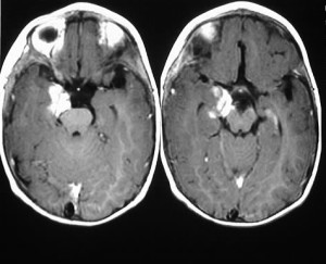
Gangliogliomas are partly diagnosed with techniques that provide pictures of the brain. One such technique is a CT (computerized tomography) scan. CT scanning is an advanced imaging technique that uses x-rays and computer technology to produces more clear and detailed pictures than a traditional x-ray.
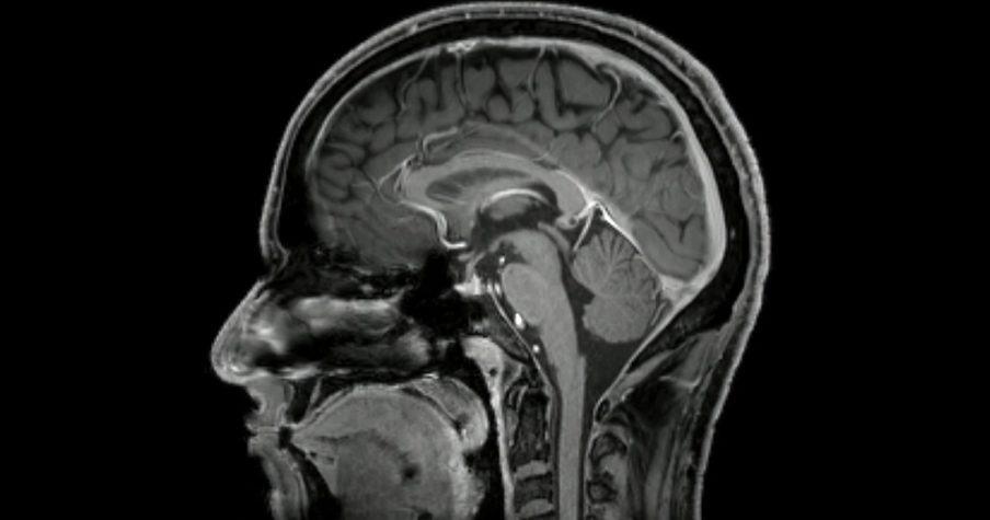
Magnetic resonance imaging (MRI) scans of the brain are often used. MRI scans produce extremely detailed pictures of the inside of the body by using very powerful magnets and computer technology. MRI scans are more detailed and more expensive that CT scans. MRI scans are the most sensitive and specific at detecting that a tumor is present, particularly when contrast is injected into the person’s body during the scan. Contrast is a substance that allows damaged areas to show up better on the picture produced by the MRI. About 50% of gangliogliomas will be detected when contrast is used.
MRI exams are performed at over 20 Capitol Imaging Services locations. Click here to learn more about MRI and their various imaging technologies. Capitol Imaging Services is doctor trusted and patient preferred.
