
CT (Computed Tomography) scans and MRI (Magnetic Resonance Imaging) scans both provide diagnostic images of the inside of your body. However, they accomplish this important task in very different ways. Here are a few key differences between CT and MRI scans.
The use of radiation
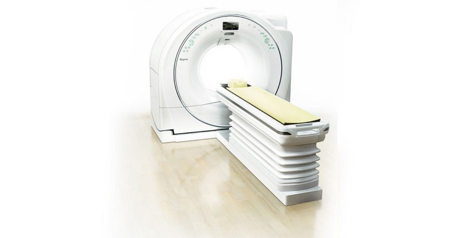
CT uses x-ray technology to produce diagnostic images. These x-rays require a small dose of ionizing radiation.
The CT scanner rotates on an axis, taking multiple two-dimensional images of a person’s body from different angles. When a computer places all of these cross-sectional images together on a monitor, the result is a three-dimensional (3D) image of the inside of the body that can reveal the presence of disease or injury to a physician.
MRI scans work an entirely different way
Instead of using ionizing radiation, MRI uses radio waves and powerful magnets to produce diagnostic images. An MRI scanner can apply a magnetic field that lines up all of your body’s protons.
Radio waves are applied to these protons in short bursts, which in turn relate a signal that is picked up by the MRI scanner. A computer processes this signal and generates a 3D image of the segment of the body being examined.
The length of time needed to complete the scan
CT scans typically take diagnostic images much more rapidly than MRI scans. A CT scan can often be completed in less than 5 minutes while most common MRI studies take an average of 30 minutes. More complex studies, particularly those involving the use of a contrast dye, will require a scan time of about 45-60 minutes to complete.
Recommended for different reasons
Advanced imaging exams are also used for different purposes, although either can be used in some instances. CT scans are extremely helpful in diagnosing serious injuries to the head, chest, abdomen, spine and pelvis, especially fractures.
The location of benign and malignant tumors are also best imaged via CT.
Conversely, MRI often does a better job of diagnosing issues in your soft tissues, joints, tendons and ligaments. Doctors frequently recommend an MRI to scan the brain, spine, neck, breast, abdomen and muscles.
MRI is considered especially good for evaluating the spine and spinal ligaments.
Capitol Imaging Services has always been aware of the importance of patient safety in medical imaging. When a CT is most appropriate, We offer people and their medical providers the option of an ultra-low dose CT exam which is performed at select Capitol Imaging Services facilities. This is simply the safest CT available within an outpatient imaging setting. In addition, all CT scans use dose modulation software to deliver the absolute minimum radiation possible in order to perform a quality study for the radiologist.
In addition, during a CT, the person undergoing the exam will be provided with maximum shielding so that only the area of the body needed for the test is scanned.
Capitol Imaging Services has been a supporter of the national Image Gently and Image Wisely campaigns for safety in medical imaging. When you choose us for any exam, please know that we will do everything possible to keep you safe and relaxed during your visit.
Click the REQUEST AN APPOINTMENT button (above right) to send us an email requesting assistance. A Capitol Imaging Services associate will contact you to arrange your visit.
Choose independent and choose Capitol Imaging Services. We are doctor trusted and patient preferred.
