

Here is a brief excerpt from an answer to the question posed above, provided by Paul Christo, M.D., Director, Pain Treatment Center, Johns Hopkins Hospital and Health System.
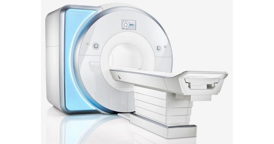
An MRI, or magnetic resonance image, is a useful tool in evaluating the spine.
An MRI is non-invasive, does not use radiation, and provides good visualization of the spinal ligaments, a herniated disc, bony infection of the spine or disc, a tumor, and spinal cord compression or damage. You cannot have an MRI if you already have a cardiac pacemaker or certain types of aneurysm clips, for example.
A CAT (CT) scan, or a computed tomography scan, uses radiation to evaluate spinal abnormalities. For instance, a CT scan can be used to evaluate spinal fractures, disc herniation, and spinal stenosis, or narrowing of the spinal canal. CT scanning is more rapid than an MRI, and provides better detail of the bones of your spine.
If your physician believes that your painful symptoms may derive from an infection or tumor in the spine, he or she may order an MRI. Either an MRI or a CAT scan is used to help diagnose disc herniation and spinal stenosis as the reason for your pain.
MRI and CT exams are performed throughout the Capitol Imaging Services affiliate network of screening and diagnostic testing centers. Our facilities are accredited in both imaging technologies by the American College of Radiology, meeting or exceeding the standards set for patient safety, technology and expertise.
Capitol Imaging Services is the largest independent radiology practice of its type serving the Southeastern United States. Not only do we perform the scan, such as a CT or MRI, we provide the radiology expertise to review test images and issue critical findings to physicians and other health care providers.
Click the green REQUEST AN APPOINTMENT button (above right) to send us an email requesting assistance. A Capitol Imaging Services associate will contact you to arrange your visit.
Capitol Imaging Services is doctor trusted and patient preferred.
