
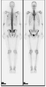
Bone Imaging is used to detect skeletal lesions as the earliest possible time, monitor the course of skeletal diseases and evaluate the metabolic activity of skeletal lesions. It can also detect osteomyelitis as well as stress fractures.
 The injection given is called 99mTc Medronate (MDP), which is safely absorbed in the bones and will localize in the tissue or organ desired. In this case, the skeletal system is the site for absorption. 99% of the calcium in our bodies are found in the skeletal system. Calcium is absorbed in the gut and continuously lost from the body through renal excretion, fecal loss and sweat. Therefore, after a nuclear medicine bone scan, we ask that you drink plenty of water to excrete the remainder of the medicine out of your body through urination.
Upon injection through a vein, the MDP medication is now traveling through your circulatory system. Over that 2-3 hour wait, the dose is absorbing through the calcium channels of all of the bones in your whole body! The bottom line for why we have you wait: this time period will ensure optimal images for diagnostic purposes.
If we were to start scanning immediately after injection, the image would have too much background tissue uptake and we would not be able to see the defined outline of the skeletal system.
Nuclear medicine studies are performed at select Capitol Imaging Services affiliate centers. Click here to learn more about the common nuclear medicine tests we perform.
Capitol Imaging Services is the largest independent radiology practice of its type serving the southeastern United States. Not only do we perform the scan such as a CT or MRI, we provide the radiology expertise to review test images and issue important results to physicians and other health care providers.
Capitol Imaging Services is doctor trusted and patient preferred.
