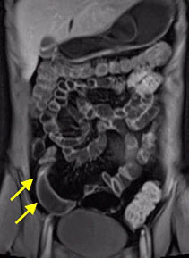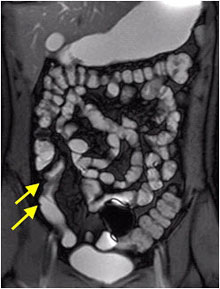
 MR enterography is a special type of magnetic resonance imaging (MRI) performed with a contrast material to produce detailed images of the small intestine.
Physicians use MR enterography to identify and locate:
- the presence of and complications from Crohn’s disease and other inflammatory bowel diseases
- inflammation
- bleeding sources and vascular abnormalities
- tumors
- abscesses and fistulas
- bowel obstructions
The benefits of Enterography with MRI are:
- MRI is a noninvasive imaging technique that does not involve exposure to ionizing radiation.
- MRI enables the discovery of abnormalities that might be obscured by bone with other imaging methods.
- The contrast material used in MRI exams is less likely to produce an allergic reaction than the iodine-based contrast materials used for conventional x-rays and CT scanning.
- MR enterography is a complementary imaging examination that helps identify areas of bowel inflammation due to such diseases as Crohn’s.
- MR enterography may eliminate the need for video capsule endoscopy (VCE).
Because MR enterography does not involve ionizing radiation, the procedure may be preferred for the evaluation of young patients with inflammatory bowel disease who may undergo multiple exams throughout their life.
If you, or someone you know, faces multiple exams due to bowel disease or Crohn’s, MR Enterography is an excellent alternative.

