
Mammography
 In women's health, regular screening for breast cancer should be top of mind. This is done at Capitol Imaging Services (CIS) with mammography.
A mammogram is an x-ray picture of the breast. There are two types of mammograms: a screening mammogram and a diagnostic mammogram.
Screening mammograms are routinely administered to detect breast cancer in women who have no apparent symptoms. Diagnostic mammograms are used to diagnose unusual breast changes, such as a lump, pain, nipple discharge, change in breast size or shape or previous breast cancer. If your screening mammogram does show an abnormality, you may need additional imaging such as a diagnostic mammogram, breast ultrasound and/or a breast MRI.
To supplement mammography, CIS incorporates digital Computer-Aided Detection (CAD), which highlights characteristics commonly associated with breast cancer. When activated, it flags abnormalities to help the radiologist detect early breast cancer. CAD is, in essence, a second set of eyes to support and enhance the radiologist’s judgment. CAD has been shown to increase breast cancer detection rates by 15 percent and up to 15 months earlier, is a part of the standard procedure for reading mammogram results.
Breast cancer screening tools at CIS includes an advanced form of imaging known as 3D mammography or breast tomosynthesis. 3D mammography gives radiologists the ability to view inside the breast layer by layer, helping to see the fine details more clearly by minimizing overlapping tissue.
Mammography also incorporates the Tyrer-Cuzick risk assessment model. The model, a mathematical tool, uses different personal and extensive family risk factors to calculate a breast cancer risk percentage of average, intermediate or high risk. This risk assessment is automatically included on each CIS screening mammogram report.
When would I get a Mammogram?
Breast cancer screening mammograms are often recommended on an annual basis, usually performed one year and one day after the previous screening. It can help reduce the number of deaths from breast cancer among women ages 40 to 70.
Mammograms are also recommended for younger women who have symptoms of breast cancer or who have a high risk of the disease.
What Will I Experience?
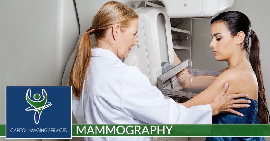
Our technologist will position your breast in the mammography unit. Your breast will be placed on a special platform and compressed with a clear plastic paddle. The technologist will gradually compress your breast.
You will be asked to change positions between images. The routine views are a top-to-bottom view and an angled side view. The process will be repeated for the other breast.
You will feel pressure on your breast as it is squeezed by the compression paddle. Some women with sensitive breasts may experience discomfort.
There are a variety of reasons that breast compression is necessary for a mammogram. Breast thickness needs to be evened out so that all tissue can be visualized. Spreading out breast tissue will minimize the likelihood that small abnormalities will be hidden by overlying breast tissue. Compression holds the breast in place to minimize any blurring that could result from motion.
A screening mammogram will take approximately 10 minutes to complete. A diagnostic mammogram may take up to 30 to 45 minutes to complete.


