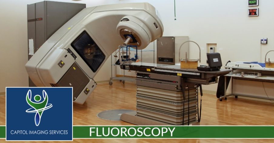
Fluoroscopy is a study of moving body structures. A continuous x-ray beam is passed through the body part being examined. The beam is transmitted to a monitor so that the body part and its motion can be seen in detail. Fluoroscopy, as an imaging tool, enables physicians to look at many body systems, including the skeletal, digestive, urinary, respiratory and reproductive systems.
Fluoroscopy may be performed to evaluate specific areas of the body, including the bones, muscles and joints, as well as solid organs, such as the heart, lung or kidneys.
Other related procedures that may be used to diagnose problems of the bones, muscles, or joints include x-rays, Computed Tomography (CT) scan, Magnetic Resonance Imaging (MRI) and arthrography.
When would I undergo Fluoroscopy?
Fluoroscopy would be considered an appropriate imaging technique for tests such as:
- Barium enema
- Barium swallow
- Hysterosalpingography
- Small bowel follow-through
- Upper GI exam
- Intravenous Pyelogram or IVP
Other uses of fluoroscopy include:
- locating foreign bodies
- visualization of a joint or joints for arthrography procedures
- image-guided anesthetic injections into joints or the spine.
What Will I Experience?
A contrast substance may be given, depending on the type of procedure that is being performed, via swallowing, enema, or an intravenous (IV) line in your hand or arm. You will be positioned on the x-ray table. Depending on the type of procedure, you may be asked to assume different positions, move a specific body part or hold your breath at intervals while the fluoroscopy is being performed.
A special x-ray machine will be used to produce the fluoroscopic images of the body structure being examined or treated. A dye or contrast substance may be injected into the IV line in order to better visualize the organs or structures being studied.
In the case of arthrography (visualization of a joint), any fluid in the joint may be aspirated (withdrawn with a needle) prior to the injection of the contrast substance. After the contrast is injected, you may be asked to move the joint for a few minutes in order to evenly distribute the contrast substance throughout the joint.
While fluoroscopy itself is not painful, the particular procedure being performed may have some discomfort, such as the injection into a joint or accessing of an artery or vein. In these cases, the radiologist will take all comfort measures possible, which may include local anesthesia or mild sedation.


