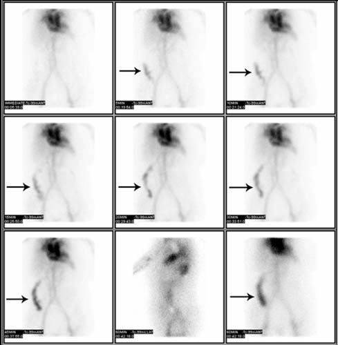
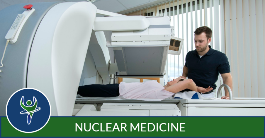
Nuclear Medicine is an imaging technology that involves the use of small amounts of radioactive materials (or tracers) to help diagnose and treat a variety of diseases. Nuclear medicine determines the cause of the medical problem based on the function of the organ, tissue or bone.
Nuclear medicine diagnostic imaging procedures are noninvasive and, with the exception of intravenous injections, are usually painless medical tests that help physicians diagnose and evaluate medical conditions. In many centers, nuclear medicine images can be superimposed with Computed Tomography (CT) or Magnetic Resonance Imaging (MRI) to produce special views, a practice known as Image Merge or Image Fusion. These views allow the information from two different exams to be correlated and interpreted on one image, leading to more precise information and accurate diagnoses.
In addition, Single Photon Emission Computed Tomography/Computed Tomography (SPECT/CT) and Positron Emission Tomography/Computed Tomography (PET/CT) units perform both imaging exams at the same time.

When Would I be Recommended for a Nuclear Medicine Test?
There are numerous applications in which a nuclear medicine study would be considered an appropriate imaging technique, such as the following:
- stage cancer by determining the presence or spread of cancer in various parts of the body
- analyze native and transplant kidney function
- scan lungs for respiratory and blood flow problems
- evaluate bones for fractures, infection and arthritis
- investigate abnormalities in the brain, such as seizures, memory loss and abnormalities in blood flow.
What Will I Experience?
You will lie on an examination table. If necessary, a technologist will insert an intravenous (IV) catheter into a vein in your hand or arm.
Depending on your type of nuclear medicine exam, the radiotracer is injected intravenously, swallowed or inhaled as a gas.
It can take anywhere from several seconds to several days for the radiotracer to travel through your body and accumulate in the area under study. As a result, imaging may be done immediately, a few hours later, or even several days after you receive the radioactive material.
When it is time for the imaging to begin, the camera or scanner will take a series of images. The camera may rotate around you or it may stay in one position and you may be asked to change positions in between images. While the camera is taking pictures, you will need to remain still for brief periods of time. In some cases, the camera may move very close to your body. This is necessary to obtain the best quality images. If you are claustrophobic, you should inform the technologist before your exam begins.
If a probe is used, this small hand-held device will be passed over the area of the body being studied to measure levels of radioactivity. Other nuclear medicine tests measure radioactivity levels in blood, urine or breath.
The length of time for nuclear medicine procedures varies greatly, depending on the type of exam. Actual scanning time for nuclear imaging exams can take from 20 minutes to several hours and may be conducted over several days.


