

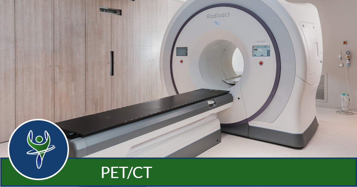
PET/CT
PET/CT combines two imaging technologies to provide information about the location and metabolism of cancer cells to aid in diagnosis and treatment. The first, PET, or Positron Emission Tomography, is a nuclear medicine study that detects the metabolic signals given off by cancer cells growing in the body.
With a PET study, a tiny amount of a radioactive substance, called a radionuclide, is used to assist in the examination of the tissue under study. PET will evaluate the metabolism of a particular organ or tissue, so that information about the physiology, or functionality, and anatomy, or structure, of the organ or tissue is evaluated.
The result is PET may detect biochemical changes in an organ or tissue that can identify the onset of a disease process before anatomical changes related to the disease can be seen with other imaging processes.
The biochemical changes are detected with the second technology, CT, which uses x-rays to locate the precise location of these changes which may indicate cancerous cells, also know as lesions or tumors.
When would I get a PET/CT Scan?
Physicians often use the PET/CT examination to:
- differentiate malignant from non-malignant disease (diagnosis)
- determine whether a cancer has spread in the body (staging)
- assess the effectiveness of a treatment plan, such as cancer therapy (response to therapy)
- determine if a cancer has returned after treatment (restaging).
What Will I Experience?
Nuclear medicine diagnostic imaging procedures are noninvasive and, with the exception of intravenous injections, are usually painless medical tests that help physicians diagnose and evaluate medical conditions. These imaging scans use radioactive materials called radiopharmaceuticals or radiotracers.
The radiotracer is injected into the body and takes about 30-60 minutes to travel through your body and be absorbed by the area under examination. Radioactive emissions from the radiotracer are detected by a special camera or imaging device that produces pictures and detailed molecular information.
You will lie flat on the examination table. If necessary, a technologist will insert an intravenous (IV) catheter into a vein in your hand or arm. You may be asked to drink some contrast material that will localize in the intestines and help the radiologist interpreting the exam.
When you are moved into the PET/CT scanner to begin the exam, the CT exam is performed first, followed by the PET scan. There may be an occasion when a second CT scan follows the PET imaging.
Typically, a PET/CT exam takes approximately 30 minutes to complete.

