
Echocardiogram
How is an Echocardiogram Created?
When Would I get an Echocardiogram?
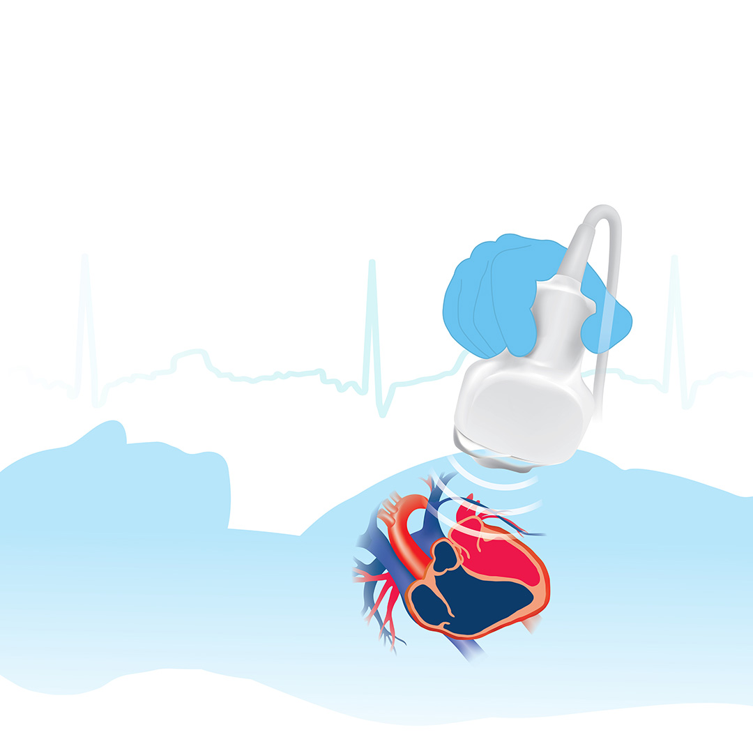
An echocardiogram creates detailed images of your heart’s chambers, valves, walls and the blood vessels, known as the aorta, arteries and veins, attached to your heart.
Your medical provider may recommend an echocardiogram if:
- you have a heart murmur
- you have had a heart attack
- you have unexplained chest pains
- you have had rheumatic fever
- you have a congenital heart defect.
This exam is designed to provide information on:
- the size and shape of your heart
- the size, thickness and movement of your heart’s walls
- the heart’s pumping strength
- if the heart valves are working correctly
- if blood is leaking backwards through your heart valves, known as regurgitation
- if the heart valves are too narrow, known as stenosis
- if there is a tumor or infectious growth around your heart valves.
An echocardiogram can also identify problems with the outer lining of your heart, known as the pericardium, with large blood vessels entering and leaving the heart, blood clots in your heart’s chambers or abnormal holes between the chambers.
What Will I Experience?
You will lie on your back on an exam table. You may have your position adjusted to either side in order to improve the quality of the images captured during the exam. The technologist will place small metal disks, called electrodes, on your chest. The disks have wires that hook to an electrocardiograph machine. This machine keeps track of your heartbeat during your test.
An ultrasound technologist will apply a warm water-based gel to the area of the body being studied. The gel will help the transducer make secure contact with the body and eliminate air pockets between the transducer and the skin that can block the sound waves from passing into your body. The transducer is placed on the body and moved back and forth over the area of interest until the desired images are captured.
There is usually no discomfort from pressure as the transducer is pressed against the area being examined. However, if scanning is performed over an area that is tender or sensitive, you may feel some pressure or minor discomfort from the transducer.
Once the exam is complete, the clear ultrasound gel will be wiped off your skin. Any portions that are not wiped off will dry quickly. The ultrasound gel does not usually stain or discolor clothing.
Typically, an echocardiogram takes approximately 60 minutes to complete.
