
Galactography
 Galactography, also known as a galactogram, combines mammography with an injection of contrast material to create images of the inside of a woman’s breast’s milk ducts. It is most commonly used when a woman has experienced a bloody or clear discharge from the breast nipple, but has an otherwise normal screening mammogram.
It’s important not to squeeze the nipple prior to the exam as there may only be a small amount of fluid and it is necessary to see where it is coming from to perform the exam.
A mammogram is an x-ray picture of the breast.
When would I get Galactography?
The most common use of galactography is to evaluate a woman who has a bloody or clear discharge from her breast nipple and an otherwise normal mammogram.
Galactography is typically NOT called for in women with the following conditions:
- a discharge that is milky, blue-green, green, or gray is usually not a cause for concern, especially if it comes from multiple ducts in the breast
- a discharge that is from both breasts in a woman who has not had children may indicate a side effect from a drug, or may be related to a pituitary problem located in the brain.
If there is a filling defect, known as a black area, in the milk duct, it often indicates a small mass. Most of these are papillomas, which are non-cancerous masses of the milk ducts. They may be precancerous, and sometimes are removed. Less than 10 percent of filling defects will be cancer.
The galactogram will not only find the small mass, but will also show where it is located in the breast, to help the radiologist find the area.
What Will I Experience?
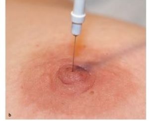
In galactography, a small amount of contrast material is injected into the milk duct, and a mammogram is performed so that the inside of the milk duct can be seen.
The patient is seated or placed on her back with the breast exposed. The nipple is cleansed, and a tiny amount of fluid is squeezed from the nipple to identify the duct with the discharge. The milk duct may be dilated to permit a small catheter (a plastic, hollow tube) or blunt-tipped tube to be inserted into the milk duct.
Occasionally, a warm towel will be placed on the breast to help the milk duct become more visible and to allow easier access to the milk duct. A small amount of contrast material is then injected and a mammogram is obtained. A second injection and mammogram may be performed.
You must hold very still and may be asked to keep from breathing for a few seconds while the x-ray picture is taken to reduce the possibility of a blurred image. The technologist will walk behind a wall or into the next room to activate the x-ray machine.
When the examination is complete, you may be asked to wait until the radiologist determines that all the necessary images have been obtained.
Typically, a galactogram takes approximately 30 to 60 minutes to complete.

