Inflammatory breast cancer (IBC) is a rare, aggressive cancer that requires a timely diagnosis, since the five-year survival rate is just 25-50 percent. Used in combination, PET and CT scans provide faster, more comprehensive detection of systemic spread for IBC patients.
IBC accounts for between 1-5 percent of all breast cancers in the United States, according to Radiology Today. But cancer in initially diagnosed IBC patients has already metastasized about 20 percent of the time, approximately doubling the percentage of other breast cancer patients.
Oftentimes, this is because inflammatory breast cancer presents with unique symptoms. IBC patients experience rapid swelling, changing skin texture, induration, tenderness, and warmth. Swelling and redness occur when cancer cells block the skin’s lymph vessels.
How Do PET/CT Scans Work?
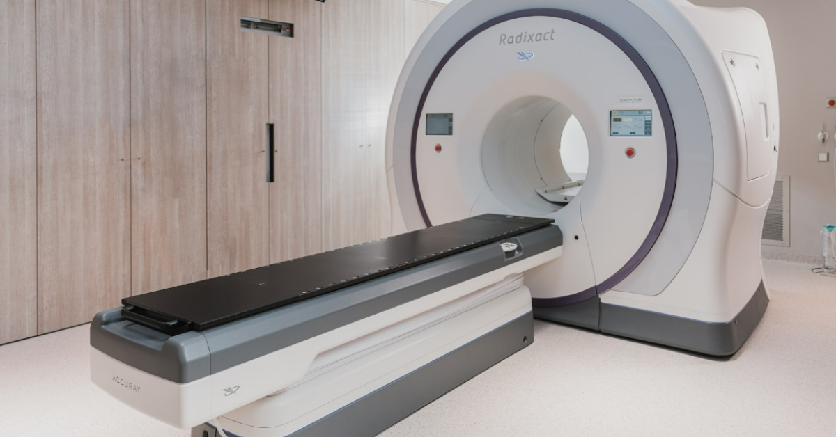
PET and CT scans are two imaging processes that help doctors learn the location and metabolism of cancer cells in order to make proper diagnosis and treatment plans.
Computerized tomography (or a CT scan) combines images from different angles to create cross-section pictures of the body with more information than typical X-ray technology. Positron Emission Tomography (a PET scan) uses nuclear medicine to detect metabolic signals emitted by cancer cells.
A tiny amount of a substance called a radionuclide is injected during a PET scan. This tracer travels through the blood system to tissue and organs, allowing a radiologist to pinpoint problem areas. The CT scan then provides a more complete picture of the changes, including possible lesions or tumors.
Why is PET/CT Critical in Diagnosing IBC?
IBC patients aren’t always diagnosed with a palpable mass, so it’s difficult to target a biopsy. At the same time, IBC is defined as a systemic disease, meaning it can rapidly spread through bone marrow or lymph nodes – so time is of the essence.
PET/CT imaging was confirmed to improve survival rates for IBC patients, according to a recent study by the University of Texas MD Anderson Cancer Center presented at the American Roentgen Ray Society’s annual meeting. PET and CT scans have long been used for oncology imagining, but only more recently have they been combined to help doctors more easily detect cancers.


