

Cardiac MRI
Magnetic Resonance Imaging (MRI) of the heart—often called cardiac MRI—uses a powerful magnet and radio waves to create detailed pictures of your heart and nearby blood vessels. These images help doctors evaluate heart structure and function, look for inflammation or scarring, and guide treatment plans without using ionizing radiation.
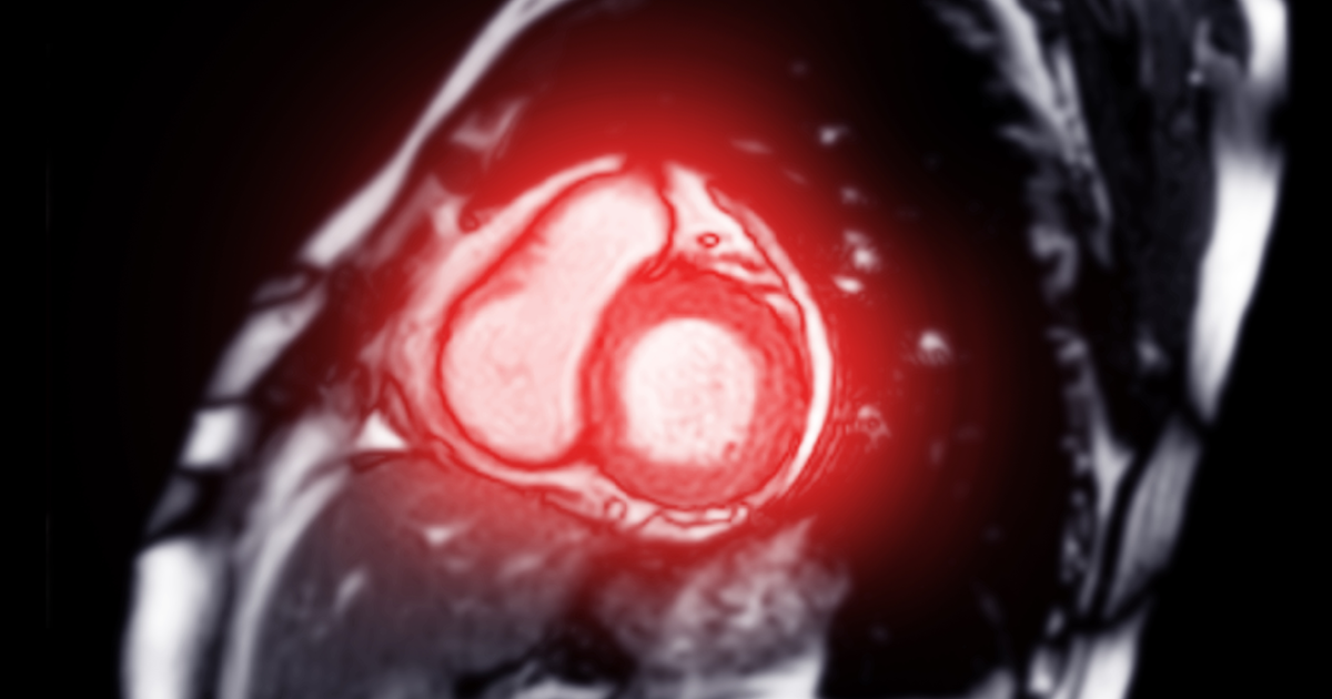
Cardiac MRI can show how well your heart muscle squeezes, how blood flows through the chambers and valves, and whether there are signs of prior heart injury (such as scar from a past heart attack) or conditions like myocarditis, cardiomyopathy, or pericardial disease. It is also useful in congenital (from birth) heart conditions for both children and adults.
When would I get a Cardiac MRI?
Your medical provider may recommend a cardiac MRI to help evaluate or monitor:
- Unexplained chest pain, shortness of breath, fainting, or abnormal heart rhythms
- Cardiomyopathy (weak or thickened heart muscle), myocarditis (heart muscle inflammation), or pericarditis (inflammation around the heart)
- Prior heart damage or scarring from a heart attack and to assess heart muscle viability
- Congenital heart disease (before or after treatment)
- Valve problems, pulmonary hypertension, or diseases of the aorta and great vessels
- Planning or tracking the effects of cardiac treatments or surgery over time
What Will I Experience?
Cardiac MRI exams are noninvasive and painless. Most take about 30–90 minutes once imaging starts. You’ll lie on a padded table that slides into the scanner; a technologist can see, hear, and speak with you at all times through a two-way intercom. You’ll receive earplugs or headphones for the knocking or thumping sounds the scanner makes. Many exams include short breath-holds so pictures won’t blur.
Small electrocardiogram (ECG) patches are usually placed on your chest to help time the pictures with your heartbeat. It’s common to feel the imaged area become slightly warm. Please stay as still as possible so we can capture clear images. If you feel anxious or claustrophobic, talk with your provider—mild sedation can be arranged when appropriate. If you receive a sedative, you’ll need someone to drive you home.
Contrast and Cardiac MRI
Preparation for MRI
- Clothing and metal: You may change into a gown. Remove jewelry, piercings, watches, credit cards, and electronic devices before the scan; metal can affect image quality and is unsafe near the magnet.
- Eating and medicines: Unless told otherwise, take your usual medicines and eat normally. If stress imaging is planned, follow any no-caffeine instructions you receive.
- Implants and devices: Tell us about any implanted devices (such as pacemakers, defibrillators, aneurysm clips, cochlear implants, infusion pumps, or metal fragments). Many modern cardiac devices are MRI-conditional and can be scanned under specific safety protocols, but we must confirm details in advance.
- Pregnancy: MRI has been used for decades with no known harmful effects; however, contrast is avoided during pregnancy unless clearly necessary. Please tell your technologist if you are or might be pregnant.

