

A person visited Capitol Imaging Services due to experiencing low back and right leg pain. They also had a previous medical history of lumbar spine surgery.
In this particular instance, the medical provider recommended a Computerized Tomography (CT) scan that was part of a specialty nuclear medicine Image Merge study in which the two imaging techniques, CT and nuclear medicine, were combined to form a more complete picture of their anatomy.
Pars Fracture
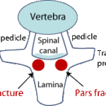 One of the clinical questions the medical provider wanted to have answered was if there was a Pars fracture. According to the Palo Alto Medical Foundation, a Pars fracture:
- is the most common cause of lower back pain in adolescent athletes.
- usually causes low back pain that is predominantly on one side of the back, as opposed to in the center of the back.
- is caused by overuse and usually starts as mild pain that gradually worsens with running, jumping and kicking activities.
- is usually felt most while arching backward, rotating at the waist or straightening up from bending forward.
- typically gets worse with sports and better with rest.
Pars stress fractures involve a small connecting bone in the lumbar spine, called the pars interarticularis. The pars bone is a small bone that connects the facet joints that are a chain of joints found on each side of the spine. The facet joints spread apart and have no pressure on them when you are sitting or bending forward, but they press against each other and are under pressure during activities such as running, jumping, kicking, rotating or arching backward.
These stress fractures are not visible on x-rays of the spine until they have been present for many months. By the time they can be seen on x-rays, they do not have the ability to heal. Most athletes seek medical evaluation because of significant pain at a point before the stress fracture is visible on an x-ray.
The stress fracture can be diagnosed with an MRI, CT or bone scan.
Capitol Imaging Services is an independent radiology practice that can bring together multiple imaging techniques in order to provide this complete diagnostic picture. We can merge the anatomical imaging of CT or MRI with the functional imaging of nuclear medicine. Capitol Imaging Services provides the radiology expertise to review test images and issue important findings to medical subspecialists, who benefit from both studies separately analyzed and then reported on together.
CT, MRI and bone scan studies are performed at multiple Capitol Imaging Services locations. That’s a major reason why Capitol Imaging Services is doctor trusted and patient preferred.
Click the REQUEST AN APPOINTMENT button (above right) to send us an email requesting assistance. A Capitol Imaging Services associate will contact you to arrange your visit.
