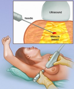
 Lumps or abnormalities in the breast are often detected by physical examination, mammography, or other imaging studies. However, it is not always possible to tell from these imaging tests whether a growth is benign or cancerous.
A breast biopsy is performed to remove some cells—either surgically or through a less invasive procedure involving a hollow needle—from a suspicious area in the breast and examine them under a microscope to determine a diagnosis. Image-guided needle biopsy is not designed to remove the entire lesion, but most of a very small lesion may be removed in the process of biopsy.
Image-guided biopsy is performed by taking samples of an abnormality under some form of guidance such as ultrasound, MRI or mammographic guidance.
In ultrasound-guided breast biopsy, ultrasound imaging is used to help guide the radiologist’s instruments to the site of the abnormal growth.
Capitol Imaging Services performs ultrasound-guided breast biopsies. By being performed in an independent outpatient setting, women experience a more friendly, relaxed and comfortable environment and experience first-class service from our staff. Because a woman chose to have her biopsy at Capitol Imaging Services, she avoided the crowded, noisy and hectic hospital and their mega-high costs and charges.

