
CT of the Chest
Computed Tomography, or CT of the chest, combines x-rays with computer technology to create detailed images of bones, tissues, and organs in the chest. Unlike standard x-rays that capture a single image of the entire chest, CT scans provide images in "slices," allowing for a more detailed view.
In standard x-rays, dense tissues like bones can obscure the view of other body parts. However, in a chest CT scan, the various image slices clearly show both bones and underlying soft tissues. This helps physicians diagnose and detect many chest conditions at an early stage.
The procedure is short, painless, and emits very low amounts of radiation.
When would I get a CT of the Chest?
Your medical provider may recommend a CT of the chest to:
- Examine abnormalities found on conventional chest x-rays
- Help diagnose the causes of symptoms such as cough, shortness of breath, chest pain, or fever
- Detect and evaluate the extent of tumors in the chest or those that have spread from other parts of the body
- Assess whether tumors are responding to treatment
- Help plan radiation therapy
- Evaluate injuries to the chest, including the heart, blood vessels, lungs, ribs, and spine
- Evaluate abnormalities of the chest found on fetal ultrasound examinations
A chest CT may also be utilized to evaluate acquired and congenital lung abnormalities such as pneumonia, interstitial lung disease or tumor evaluation.

What Will I Experience?
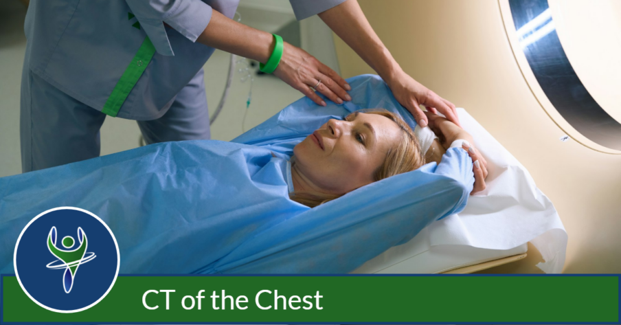
During a CT scan of the chest, the patient is positioned on a table that moves inside a ring or “gantry” that is part of the CT scanner. As the table moves, the x-ray scanning devices within it rotate around your chest area to obtain the necessary views.
Most people find that the most challenging part of a CT exam is holding their breath during the scan. However, the technologist will inform you when to suspend respirations and when you can breathe normally. Typically, the actual scan time is around 20 seconds.
The appointment typically takes about 15-20 minutes, including time for the exam interview and preparation. In some cases, an intravenous contrast injection is required to highlight certain anatomical structures so the radiologist can visualize them in more detail.


