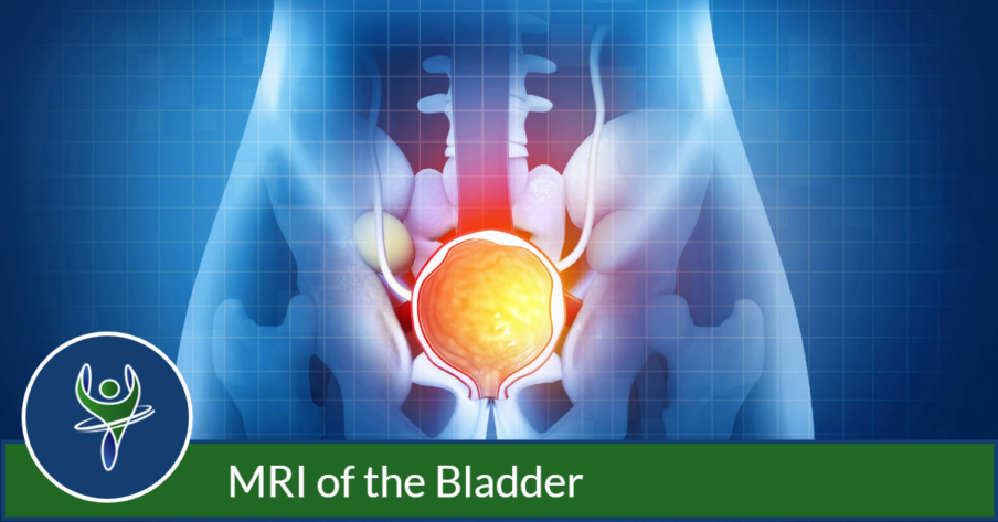
MRI of the Bladder
Magnetic Resonance Imaging, or MRI, helps doctors better understand the condition of your bladder, allowing them to potentially stage bladder cancer, rule out related issues with the urinary tract, and create unique treatment programs.
MRI scans utilize a powerful magnet to create detailed, cross-sectional pictures of the bladder. The resulting images can confirm that urinary systems are functioning normally. Should cancer be found, an MRI can also show how far the disease has progressed within the bladder or potentially spread outside of it.
MRIs have become more common because no ionizing radiation is used. They’re also preferred over CT (computed tomography) scans for some people with reduced kidney function, since patients may risk damage from prolonged exposure to the CT scan’s contrast materials.
When would I get an MRI of the Bladder?
Your medical provider may recommend an MRI of the bladder if you experience:
- Difficulty emptying your bladder
- Bladder control problems
- Urgent or more frequent urination
- Blood in the urine (known as hematuria)
- Persistent pain in abdomen or lower back
- Swelling in the abdomen
- Recurrent urinary tract infections
What Will I Experience?
MRI exams are noninvasive and pain-free. Typically, the entire process takes about 30 to 60 minutes to complete.
Negative reactions include feeling claustrophobic inside a conventional scanner, finding it difficult to remain still during the process, or being bothered by noises that the scanner emits. Some anxious or nervous patients may be offered a mild sedative. In those cases, patients will need to have someone on hand to drive them to our center and then take them home after the exam is over.
Areas of the body will typically feel slightly warmer while being imaged. It’s important that patients remain completely still while these images are being recorded. Each image typically takes only a few seconds to capture, though some may last up to a few minutes. Patients know when images are being recorded because they hear thumping or tapping sounds as the coils that generate radiofrequency pulses are activated. There will be time to relax between imaging sequences, but patients are asked to maintain their position as much as possible.
Patients are usually alone in the exam room during these scans but a technologist will be able to see, hear and speak with them at all times using a two-way intercom. Patients are also offered earplugs or a headset to reduce noise associated with MRIs, including humming or thumping noises during imaging. MRI scanning areas are air-conditioned and well-lit; some scanners even have music to listen to during the test.
With exams requiring intravenous contrast material, patients will typically feel coolness and a flushing sensation for a minute or two following the injection. The intravenous needle may cause some discomfort when it is inserted. Some experience bruising once it’s removed. There is also a very small chance of skin irritation at the site of the IV tube insertion.


