Prostate 3T MRI with CAD
Capitol Imaging Services (CIS) provides an alternative option to diagnose prostate cancer early with our Prostate 3T MRI exam service. One man in nine will be diagnosed with prostate cancer during his lifetime. MRI can play an integral role in evaluating the prostate gland.
CIS uses 3 Tesla (3T) ultra-high field strength MRI to create detailed high resolution “multiparametric” (meaning to take a large amount of different pictures of the prostate gland) cross-sectional images of the prostate gland. In addition, techniques such as MR Diffusion and MR Perfusion Imaging are routinely utilized for better detection.
Diffusion imaging provides information on the degree of cellular crowding, which is worse where cancer cells are rapidly turning over. Perfusion imaging provides a map of blood flow, which is increased in cancer cells. These advanced preoperative imaging techniques enable surgeons to more precisely target suspicious lesions for biopsy and to more accurately decide when to remove or spare the delicate neurovascular bundles in different cases.
Click here to learn more about the advantages of multiparametric MRI at Capitol Imaging Services.
These decisions significantly affect patients’ quality of life after surgery, since urinary continence and sexual potency may be affected.
Capitol Imaging Services does not use an endorectal coil. This coil is invasive and uncomfortable both physically and mentally. Instead, CIS uses a pelvic coil for a prostate MRI study for the following reasons:
- Our 3T magnets are state-of-the-art powerful systems that generate fantastic magnetic resonance image clarity without the need for that type of coil.
- Multiparametric MRI technique is equally advanced and provides sophisticated visual data on the key parameters that differentiate healthy tissue from cancer, and from benign prostate conditions.
The CIS patient-centered approach means that we want men to feel safe and comfortable during a prostate MRI exam. It is easier to relax and lie still when required to do so if one is not preoccupied and worried about rectal pressure.
Capitol Imaging Services was the first outpatient imaging center in Louisiana to use cutting-edge advanced image analysis technology with MRI to assist radiologists, urologists, radiation and medical oncologists in the fight against prostate cancer. Our combination of the technology with an academic radiology team approach is the cornerstone of the CIS advanced prostate MRI program.
This program remains unmatched in providing more information to aid in diagnostic assessment of the prostate gland.
What is the Role of CAD?
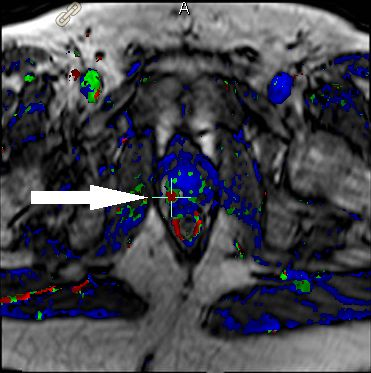
CAD is a software package designed to provide a reliable means of visualizing the presence and pattern of contrast-induced enhancement of MRI images in the prostate. This program helps prostate cancer cells stand out in situations where they might normally be undetectable.
This technology analyzes all the MRI images and creates colorized maps based on how the contrast material flows into and out of the prostate. The enhanced visual images help radiologists to distinguish benign from malignant lesions.
In the past, the prostate gland was manually outlined, which was done as part of the radiology report. With CAD analysis, our radiologists are provided with automated “advanced visualization” consisting of three dimensional (3D) images of the prostate gland. CAD will assess any lesions using PI-RADS (Prostate Imaging Reporting and Data System) scoring and incorporate the assessments into the report.
This hardware and software combination of MRI and CAD delivers the comprehensive exam evaluation required by medical providers as part of their diagnosis and potential therapy plans.